What is Melanoma?
Melanoma is a type of cancer that typically occurs in the skin but may also occur in the mouth, eyes or intestines. It develops from the melanin-forming cell known as melanocytes, hence the name.
Considered as the most dangerous form of skin cancer, melanoma stems from mutations of damaged DNA of the skin cells due to excessive exposure to ultraviolet rays from the sun or tanning beds.
Once melanoma is diagnosed, doctors determine its risk profile to develop a treatment approach. This process is called staging. The stage of a cancer describes how much the illness has spread and is used to estimate the patient's survival rate.
“If a melanoma is found on your skin and is excised/cut out, the pathologist, a specialist doctor, will tell us essentially how thick the lesion is and how deep it has invaded to the tissue we have sampled.”
“Thicker or deeper lesions are considered higher risk of spreading around the body. If this is the case, then there are other stages of melanoma which encompass whether lymph nodes have the cancer and/or other tissues in the body.” Dr Richard Barker
Determining the stage of a melanoma
There are three ways to determine the stage of a melanoma.
Microstaging
A hispatologist takes sample tissues of the tumour and surrounding areas and examines it under the microscope to find out how thick the tumour is; how deeply it has invaded the skin or connective tissues; and how widespread it is.
Clinical staging
This is a physical examination of the lymph node groups related to the affected area to see if the melanoma has spread to the lymph nodes.
Staging After Ulceration
If there is already an open sore over the melanoma, this is called ulceration. Staging after ulceration involves imaging scans such as CT, MRI and PET scans to enable doctors to see inside the body and determine whether the cancer cells have spread to the lymph nodes or other organs.
Different Stages of Melanoma
Stage 0
Stage 0 indicates the cancer cells are confined to the epidermis or outer layer of the skin and have not grown any deeper. This stage is also known as in situ, a Latin term for "in place". Treatment for this is surgical removal.
Stage I
Stage I melanoma can either be 2mm without ulceration or up to 1mm with ulceration but neither has metastases or lymph node involvement. Main treatment for this is a surgical procedure called wide local excision. If the melanoma is between 1-4mm and/or growing rapidly, removal of nearby lymph nodes may also be considered.
Stage II
Stage II melanoma is defined by tumour thickness and ulceration without evidence of metastases or lymph node involvement. The thicker the melanoma, the more chances of it spreading. Main treatment is surgical removal. Removal of nearby lymph nodes may be necessary to prevent further spread. Radiation and drug may also be required to prevent the cancer cells from recurring.
Stage III
Stage III melanoma has spread to the lymph nodes, regardless of thickness but has not metastasized. Surgical removal of the lymph nodes is usually required as well as drug treatment and radiation.
Stage IV
Stage IV melanoma has spread to regional lymph nodes and to other organs of the body such as lungs, liver, bone or brain. The thickness of tumour at this stage no longer matters. Treatments include systematic drug therapies such as immunotherapy and targeted therapy. Surgery and radiation may also be considered.
Initial Diagnosis
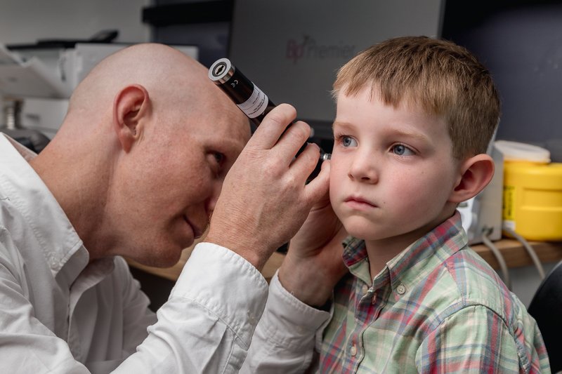
You should always take necessary steps to reduce your risk, including regular self-examinations and an annual skin check with a health professional. Knowing your own skin is crucial in detecting skin cancer early on. If you are familiar with the patterns of your moles, birthmarks, freckles, blemishes, and other marks on your skin, you can readily spot any new growths or changes in existing moles. If you find a new spot on your skin or any changes in size, you should see a doctor immediately.
Your health provider will examine all parts of your skin, including hands, feet, scalp, and even inside your nose and mouth. Should they find anything suspicious, often a biopsy is recommended. In this procedure, all or part of a mole or growth will be removed and sent to a pathologist for analysis.
After the biopsy, you will get a pathology report that contains important information that will help your doctor arrive at a diagnosis and prognosis, determine what further tests are necessary and what kind of treatment is beneficial. You should always ask for a copy of your pathology for future reference.
If you’ve had melanoma before; have dysplastic nevus syndrome; a family history of melanoma or are exposed to the sun or tanning beds excessively, it's important that you get regular skin exams. A small investment and time can prove to be a life-saving choice.